MRI NEWSLETTER: Ulnar Collateral Ligament Injury
Posted April 4, 2017
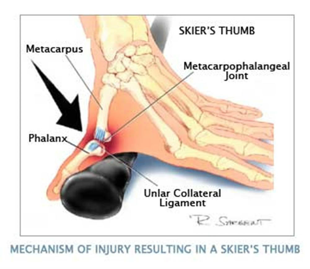
One of the most common finger injuries is a tear of the ulnar collateral ligament of the thumb at the metacarpal phalangeal joint. In the distant past, this was referred to as gamekeeper’s thumb. This was described in Scottish gamekeepers due to a repetitive injury of the ulnar collateral ligament by the gamekeepers placing stress across the MCP joint while breaking the necks of rabbits. More recently, this has been called Skier’s thumb due to the valgus stress created by falling on an outstretched hand with the thumb abducted holding a ski pole.
Physical exam and plain films are unreliable. Plain films may reveal an avulsion fracture at the base of the proximal phalanx on the ulnar side. However the fracture fragment does not reliably predict the position on of the ulnar collateral ligament, therefore MRI exam is the main stay of evaluation.
Ulnar collateral ligament injury has been divided into 4 stages by orthopedic surgeons.
Type 1 UCL Injury is a tear at base of proximal phalanx, with the UCL ligament within 3 mm of the proximal phalanx, also called an In situ injury. These injuries are treated with conservative therapy.
Type 3 UCL tear is Injury of ligament with osseous avulsion.
Type 4, the Stener Lesion. The severe hyper-abduc on of the thumb pulls the torn segment of UCL out from under the adductor pollicis longus aponeurosis. The UCL is displaced below joint space and UCL cannot contact bone to heal. Surgery is required. In complete tears of the ulnar collateral ligament, the Stener lesion occurs in 64-85% of cases, so it is very common.
Read the full article here Ulnar Collateral Ligament Injury
By Dr. William Renner
Back to News