MRI NEWSLETTER: Sinus Tarsi Syndrome
Posted June 5, 2018
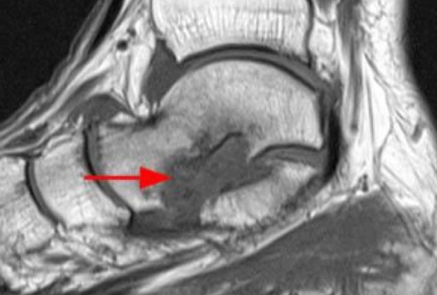
The sinus tarsi syndrome is not new, it was described by Denis O’Connor in 1958. However, the arthroscopic treatment of sinus tarsi is relatively new. The surgical treatment of sinus tarsi syndrome is arthroscopic debridement of the posterior or subtalar joint and sinus tarsi. 94% were improved at 1-8 years follow-up although half had some residual symptoms. Coronal T1 and Sagittal T2 of ankle:…
Read More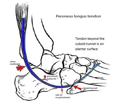
MRI NEWSLETTER: Peroneus Tendon Tears
Posted May 21, 2018
The lateral ankle tendons include the peroneus brevis (PB) and peroneus longus (PL) tendons, which serve to evert the foot. These 2 tendons share the same tendon sheath proximally at the superior peroneal retinacula (SPR) but seperate distally into two sheaths at the inferior peroneal retinacula (IPR). The brevis is located anterior to the longus. These tendons run in the retromalleolar groove on the…
Read More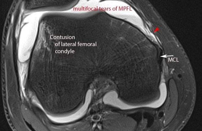
MRI NEWSLETTER: Patella Dislocation
Posted May 8, 2018
Patella disclocations are very common especially in soccer players. The history is frequently ‘rule out ACL tear’. The highest pre-test probability is females 10 to 40 years of age. The most common mechanism is sudden twisting. The classic MRI findings of lateral patella dislocation syndrome is contusion of the inferior-medial patella facet and contusion of the anterior-lateral femoral condyle. Commonly there is tearing of…
Read More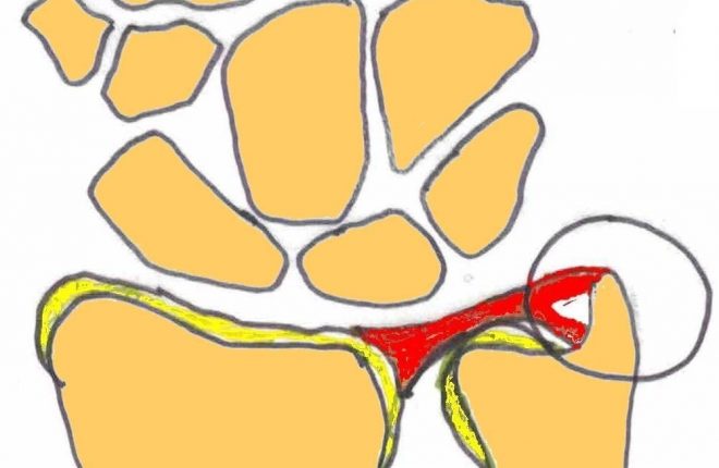
MRI NEWSLETTER: Traumatic TFC Tear
Posted May 8, 2018
Palmer divides triangular fibocartilage (TFC) tears into traumatic and degenerative. The Palmer 1B tear is a traumatic tear of the peripheral attachment of the TFC on the ulnar side with a styloid or foveal attachment (or both) tear. The foveal attachment is most important to DRUJ stability. The triangular fibrocartilage (TFC) disc is a major stabiliser of the distal radial ulnar joint. The TFC…
Read More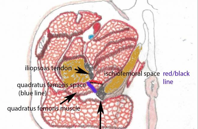
MRI NEWSLETTER: Ischiofemoral Impingement
Posted May 8, 2018
Ischiofemoral impingement is a condition that presents with pain in the hip, groin or buttock region. Pain can radiate to the lower extremity due to an irritation of the sciatic nerve. Ischiofemoral impingement is much more common in women than in men, approximately 3:1. Bilateral disease is present in one third of cases. The average patient age is older than most impingements, approximately 52…
Read More
MRI NEWSLETTER: Hip Subluxation
Posted May 8, 2018
Bo Jackson When Bo Jackson was 29 years old he was one of the most famous US athletes of all time. He is the only player to be named All-Star in both professional baseball and professional football. In 1991 while playing football against the Cincinnati Bengals he sustained a hip dislocation. He said later he reduced the hip dislocation himself. Within one year, he…
Read More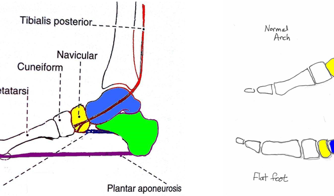
MRI NEWSLETTER: The Spring Ligament, PTT Tear and Adult Acquired Flatfoot Deformity on MRI
Posted May 12, 2017
The posterior tibial tendon is the primary stabilizer of the foot. Other stabilizers include the spring ligament, the tarsal sinus ligaments and plantar fascia. The spring ligament complex consists of the tibio-spring component of the deltoid ligament and the calcaneonavicular ligament , which limits plantar flexion of the talus and stabilizes the talocalcaneal navicular joint. The tarsal sinus ligaments (cervical and interosseous ligaments) limit […]
Read More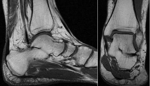
MRI NEWSLETTER: Tarsal Coalition on MR
Posted April 20, 2017
Tarsal coalition is a congenital anomaly with fusion of the tarsal bones. The fusion may be bony, fibrous or cartilaginous. The most common tarsal coalitions are the calcaneonavicular and the talocalcaneal coalition. Tarsal coalitions often lead to painful foot, frequent ankle sprain, flat foot deformity and hindfoot valgus. This may lead to peroneal spastic flat foot. Calcaneonavicular coalition ossifies early and develops symptoms at […]
Read More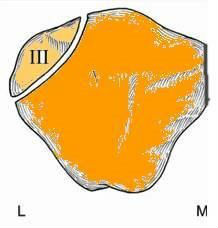
MRI NEWSLETTER: MR of the Bipartite Patella
Posted April 10, 2017
The bipartite patella is a normal patellar variant secondary to failure of fusion. Bipartite patella is often confused with a patellar fracture. Bipartite patella occurs in 8% of the population, is much more common in males 9:1, and bilateral in 50-80%. Bipartite patella occur most commonly in the superior- lateral location, 75% of cases. Bipartite patella is usually asymptomatic and found incidentally with only 2% […]
Read More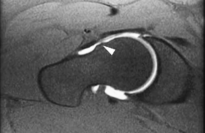
MRI NEWSLETTER: High Incidence of CAM FAI in Athletes
Posted April 4, 2017
Recent studies of FAI reveal the very high incidence of cam deformity (femoral neck bump) in Athletes. The cam deformity is a major risk factor for osteoarthritis of the hip and labral tears. More than half of the athletes studied, average age 25, had evidence of cam deformity as compared to only 23% of age-matched controls. In one study, cam deformities gradually developed during […]
Read More