News

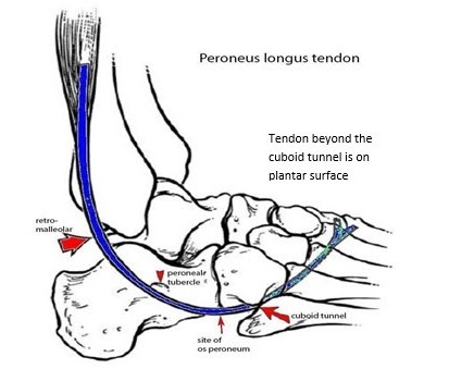
MRI NEWSLETTER: Peroneus Tendon Tears

The lateral ankle tendons include the peroneus brevis (PB) and peroneus longus (PL) tendons, which serve to evert the foot. These 2 tendons share the same tendon sheath proximally at the superior peroneal retinacula (SPR) but seperate distally into two sheaths at the inferior peroneal retinacula (IPR). The brevis is located anterior to the longus. These tendons run in the retromalleolar groove on the posterior lateral malleolus. The peroneus brevis attaches to the base of the 5th metatarsal bone. The peroneal tubercle separates the PB tendon from the PL tendon.

Peroneus brevis tears make up about 15% of ankle tendon tears; the peroneus longus tendon tears are less common and make up about 5% of ankle tendon tears.
The most frequent lateral tendon tear is a peroneus brevis tendon longitudinal spllit tear at the level of the retromalleolar groove. The PB forms the ‘chevron’ sign (an inverted V) when it tears.

Peroneus Longus (PL) Tendon:
The p. longus tendon takes 3 bends. The PL passes behind fibula in the retromalleolar region then under the peroneal tubercle, then under the cuboid to enter the cuboid tunnel to pass across the plantar aspect of the foot to insert on the plantar aspect of medial cuneiform and 1st metatarsal.
Hypertrophy of the peroneal tubercle is frequently associated with partial p. longus split tears in the middle of the peroneus longus tendon, with full thickness tears occurring in the cuboid tunnel.

Several factors may predispose to peroneal tendon injuries:
– os peroneum
– convex or flat fibular retromalleolar groove
– hypertrophy of the peroneal tubercle
– accessory peroneus quartus muscle
– low-lying peroneus brevis muscle belly

In about 20% of individuals, the peroneus longus has a sesamoid bone within the tendon, the os peroneum, which can cause pain and lead to tendon tear called the Painful Os Peroneum Syndrome or POPS. The POPS syndrome is caused by a fracture of the os, a tear of the peroneus longus tendon, or entrapment by an enlarged peroneal tubercle laterally. Peroneus brevis and longus tears are best seen on axial PD or T2 images, however an os peroneum is easiest to see on sagittal images.

Read the full article here: Peroneus Tendon Tears
By Dr William Renner

Back to News