MRI NEWSLETTER: Tarsal Coalition on MR
Posted April 20, 2017
Tarsal coalition is a congenital anomaly with fusion of the tarsal bones. The fusion may be bony, fibrous or cartilaginous. The most common tarsal coalitions are the calcaneonavicular and the talocalcaneal coalition. Tarsal coalitions often lead to painful foot, frequent ankle sprain, flat foot deformity and hindfoot valgus. This may lead to peroneal spastic flat foot. Calcaneonavicular coalition ossifies early and develops symptoms at 8-12 years of age. Talocalcaneal coalition ossifies later and develops symptoms at 12-16 years of age.
Most clinical studies suggest the prevalence of tarsal coalition to be 1% of the population however cadaver studies reveal an incidence of 13%. This would suggest that many tarsal coalitions are asymptomatic or that fibrosis or cartilaginous coalition may have fewer symptoms than bony coalition. Bony fusions are best detected by CT examination while fibrous or cartilaginous coalitions are best evaluated by MR. 50% of cases are bilateral; 20% have multiple coalitions in the same foot.
Talocalcaneal Colaition
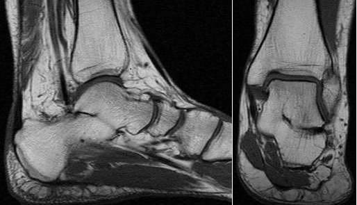
A common sign on coronal view is that the subtalar joint is no longer horizontal but has an oblique orientation. The drunker waiter sign is an abnormal angulation of the sustenaculum tail with the talus has been compared to a waiter having trouble carrying a tray, with the ‘tray’ angulated instead of horizontal.
Calcaneal Facets
The talocalcaneal coalition frequently involves the middle subtalar facet of the calcaneus at the sustentaculum tali and middle talar facet of the talus, usually involves the entire joint. Secondary signs are a hypoplastic sustentaculum tali and talar beaks.
The MR criterion for a bony coalition is a continuous marrow across the coalition. Fibrous or cartilaginous coalitions may have loss of the fat plate at the articulation. Bone marrow edema is frequently seen in the area of the coalition. If the connecting tissue is low on signal on both T1 and T2 images it is considered a fibrous coalition. Cartilaginous coalitions frequently have fluid signal at the coalition. If the bridge is gray on T2 weighted images with or without fluid signal the coalition is likely cartilaginous.
Read the full article here: Tarsal Coalition on MR
By Dr William Renner