MRI NEWSLETTER: Posterior Medial Corner Injuries of the Knee
Posted April 4, 2017
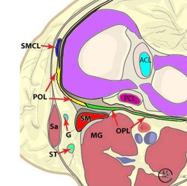
The posterior third of the posterior medial corner is made up of:
– the posterior oblique ligament (POL)
– the oblique popliteal ligament (OPL)
– the posterior third of the medial meniscus the semi-membranous tendon (SM)
The surgeons frequently ask if the POL is injured in posterior medial corner injuries.
The posterior third of the posterior medial corner is made up of:
– the semi-membranous tendon (SM)
– the posterior oblique ligament (POL)
– the oblique popliteal ligament (OPL)
– the posterior third of the medial meniscus
The posterior medial third of the knee is represented by the superficial MCL (also called tibial collateral ligament-TCL by the surgeons) and the deep MCL which is made up of the meniscofemoral and the meniscotibial ligaments. The deep MCL is separated from the superficial MCL by the MCL bursa. The superficial MCL arises from the medial femoral condyle and is a midline structure. The superficial MCL does not attach to the medial meniscus.
The posterior oblique ligament (POL) arises from the adductor tubercle, separately from the superficial MCL. The POL is angled at about 25 degrees posterior to the vertical MCL. The POL attaches to the posterior horn of the medial meniscus.
In the PCL deficient knee, the POL becomes an important posterior restraint against posterior tibial translation.
Posterior medial corner knee injuries can lead to “Anteromedial rotatory instability (AMRI)” of the knee
This is a clinical diagnosis made by the orthopedic surgeons. However the MR equivalent frequently consists of an acute ACL injury accompanied by a posterior oblique ligament (POL) injury. A pure valgus force can cause damage to the superficial MCL and the addition of rota on can lead to ACL tear.
In cases that required surgery for Anteromedial rotatory instability, injury to the POL was present in 99% of cases.
Read the full article here Posterior Medial Corner Injuries of the Knee
By Dr. William Renner
Back to News