MRI NEWSLETTER: Painful Os Peroneum Syndrome
Posted April 4, 2017
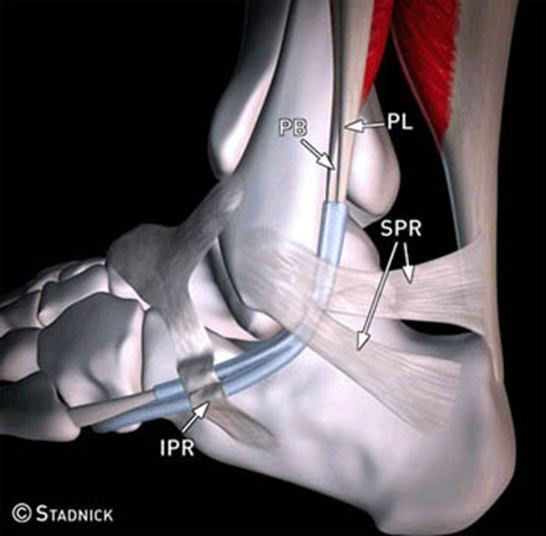
The lateral ankle tendons include the peroneus brevis (PB) and peroneus longus (PL) tendons, which serve to evert the foot. These 2 tendons share the same tendon sheath proximally at the superior peroneal retinacula (SPR), but separate distally into two sheaths at the inferior peroneal retinacula (IPR). There brevis is located anterior to the longus. These tendons run in the retromalleolar groove on the posterior lateral malleolus. The peroneus brevis attaches to the base of the 5th metatarsal bone.
Peroneus brevis tears make up about 15% of ankle tendon tears; the peroneus longus tendon tears are less common and make up about 5% ankle tendon tears.
The most frequent lateral tendon tear is a peroneus brevis tendon longitudinal split tear at the level of the retromalleolar groove. Hypertrophy of the peroneal tubercle is frequently associated with partial p. longus split tears in the middle of the peroneus longus tendon, with full thickness tears in the cuboid tunnel.
Several factors may predispose to peroneal tendon injuries:
– os peroneum
– convex or flat fibular retromalleolar groove
– hypertrophy of the peroneal tubercle at the lateral aspect of the calcaneus
– accessory peroneus quartus muscle
– low-lying peroneus brevis muscle belly
In about 20% of individuals, the peroneus longus has as a sesamoid bone within the tendon, the os peroneum, which can cause pain and lead to tendon tear called the Painful Os Peroneum Syndrome or POPS. The POPS syndrome is caused by a fracture of the Os, a tear of the peroneus longus tendon, or entrapment by an enlarged peroneal tubercle laterally. Peroneus brevis and longus tears are best seen on axial PD or T2 images, however, an os peroneum is easiest to see on sagittal images.
The osseous peroneum sesamoid may be bipartite or tripartite. A multipartite os may look like an acute fracture. An acute fracture should have irregular margins where smooth and round borders suggest a multipartite Os. Separation of the Os fragments by more than 6 mm suggest an acute fracture. If the separation is only a few millimeters, healing of a fracture or chronic impingement is more likely. A peroneus longus tendon tear may be present and is best seen as high signal on T2-weighted images. Other findings include edema of the bone marrow at the tubercle or hypertrophy of the peroneal tubercle.
Read the full article here Painful Os Peroneum Syndrome
By Dr. Bill Renner
Back to News