MRI NEWSLETTER: MR of the Bipartite Patella
Posted April 10, 2017
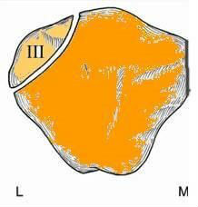
The bipartite patella is a normal patellar variant secondary to failure of fusion. Bipartite patella is often confused with a patellar fracture. Bipartite patella occurs in 8% of the population, is much more common in males 9:1, and bilateral in 50-80%. Bipartite patella occur most commonly in the superior- lateral location, 75% of cases. Bipartite patella is usually asymptomatic and found incidentally with only 2% […]
Read More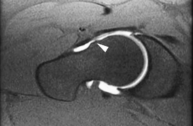
MRI NEWSLETTER: High Incidence of CAM FAI in Athletes
Posted April 4, 2017
Recent studies of FAI reveal the very high incidence of cam deformity (femoral neck bump) in Athletes. The cam deformity is a major risk factor for osteoarthritis of the hip and labral tears. More than half of the athletes studied, average age 25, had evidence of cam deformity as compared to only 23% of age-matched controls. In one study, cam deformities gradually developed during […]
Read More
MRI NEWSLETTER: Flexor Pulley Injury in Rock Climbers
Posted April 4, 2017
Injuries are most common in rock climbing and other sports resulting in a forced extension of a flexed finger. Approximately 30% of all hand injuries in rock climbers are pulley injuries. Findings on MRI include; focal discontinuity of pulley fibres, bowstringing – increased gap between the flexor tendon and volar surface of the phalanx, edema superficial and deep to pulley, fluid within tendon sheath. Read […]
Read More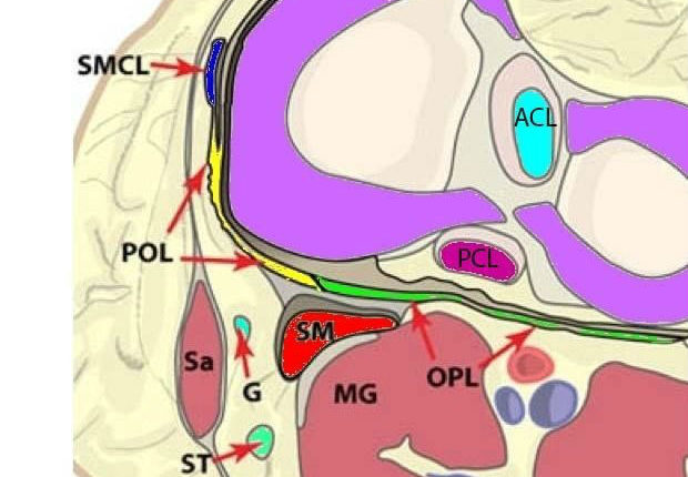
MRI NEWSLETTER: Posterior Medial Corner Injuries of the Knee
Posted April 4, 2017
The posterior third of the posterior medial corner is made up of: – the posterior oblique ligament (POL) – the oblique popliteal ligament (OPL) – the posterior third of the medial meniscus the semi-membranous tendon (SM) The surgeons frequently ask if the POL is injured in posterior medial corner injuries. The posterior third of the posterior medial corner is made up of: – the […]
Read More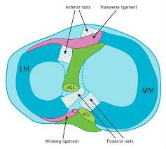
MRI NEWSLETTER: Meniscal Root Tears
Posted April 4, 2017
Meniscal root tears were not reported in the literature 10 years ago but recently have become increasingly important in both radiology and orthopedics. The meniscal roots are the primary structural anchors of both the medial and the lateral meniscus to the tibial plateau. As you can see in diagram below, the posterior roots of both the lateral and medial meniscal roots are anterior to […]
Read More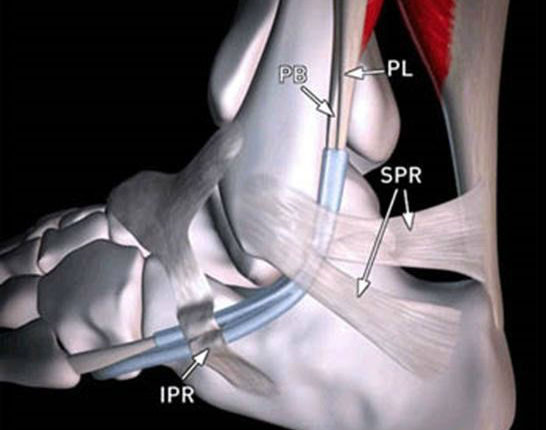
MRI NEWSLETTER: Painful Os Peroneum Syndrome
Posted April 4, 2017
The lateral ankle tendons include the peroneus brevis (PB) and peroneus longus (PL) tendons, which serve to evert the foot. These 2 tendons share the same tendon sheath proximally at the superior peroneal retinacula (SPR), but separate distally into two sheaths at the inferior peroneal retinacula (IPR). There brevis is located anterior to the longus. These tendons run in the retromalleolar groove on the […]
Read More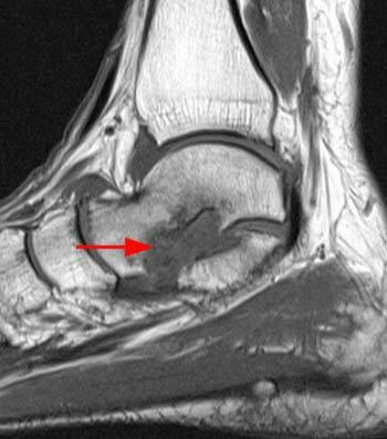
MRI NEWSLETTER Sinus Tarsi Syndrome
Posted April 4, 2017
Sinus tarsi syndrome is an inflammatory condition causing lateral hindfoot pain and instability. The most common cause for the syndrome is ankle trauma (about 70%) in particular an inversion injury, which can be treated with steroid injection into the sinus tarsi. A more serious cause of sinus tarsi syndrome is posterior tibial tendon dysfunction and spring ligament tears which frequently lead to the syndrome. […]
Read More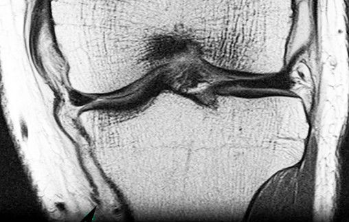
MRI NEWSLETTER: Stener-like Lesion of the Medial Collateral Ligament of the Knee
Posted April 4, 2017
An injury of the MCL is one of the most common ligamentous injuries of the knee. MCL injuries are graded 1-3, with grade 1 consisting of edema next to the MCL, Grade 2 with abnormal signal within the tendon, and Grade 3 with disruption of the tendon. Most disruptions occur at the proximal end (femoral avulsion) or mid-portion of the MCL. The vast majority […]
Read More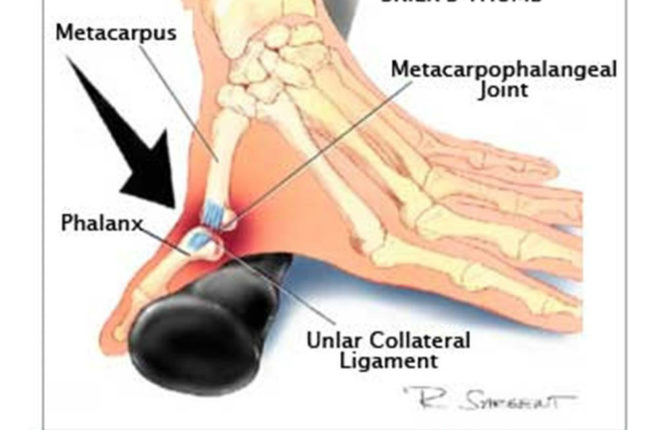
MRI NEWSLETTER: Ulnar Collateral Ligament Injury
Posted April 4, 2017
One of the most common finger injuries is a tear of the ulnar collateral ligament of the thumb at the metacarpal phalangeal joint. In the distant past, this was referred to as gamekeeper’s thumb. This was described in Scottish gamekeepers due to a repetitive injury of the ulnar collateral ligament by the gamekeepers placing stress across the MCP joint while breaking the necks of […]
Read More
MRI Intravenous Contrast Safety: UPDATE
Posted March 17, 2017
Recent media publications have outlined possible dangers of MRI contrast injections containing gadolinium and it’s potential for deposits in the human body. At Radius we use a macrocyclic contrast agent, a type which is deemed safe due to a stable chemical makeup which results in less likelihood of gadolinium being released and deposited in tissues. More information can be found here: http://www.ema.europa.eu/ema/index.jsp?curl=pages/news_and_events/news/2017/03/news_detail_002708.jsp&mid=WC0b01ac058004d5c1 MRI FAQs Do I […]
Read More