MRI NEWSLETTER: Hip Subluxation
Posted May 8, 2018
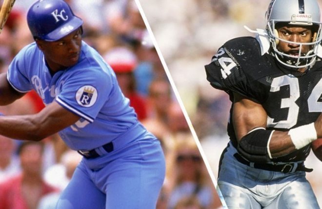
Bo Jackson When Bo Jackson was 29 years old he was one of the most famous US athletes of all time. He is the only player to be named All-Star in both professional baseball and professional football. In 1991 while playing football against the Cincinnati Bengals he sustained a hip dislocation. He said later he reduced the hip dislocation himself. Within one year, he…
Read More
Education Evenings @ Radius – Providing Great Customer Service
Posted October 1, 2017
Radius is committed to working with health practices to deliver the best possible care for patients in the region. In October our staff and guests participated in ‘Providing Great Customer Service for Healthcare Practices’, presented by Jen Flakemore. Topic – Providing great customer service for healthcare practices. – Support staff are the face of our healthcare practices. In an era of increasing competition where…
Read More
Guest Speaker – Dr William Renner
Posted August 1, 2017
In August Radius was privileged to host guest speaker Dr William Renner all the way from Cincinnati, USA. He provided a very informative and entertaining talk covering many aspects of musculoskeletal imaging, attended by Radius staff, physiotherapists and podiatrists. Dr Renner is an internationally known speaker. His lectures emphasize simple guidelines for reading MRI of the musculoskeletal system and have been praised by his students. Dr…
Read More
NeuroRAD Conference
Posted May 25, 2017
Furthering our education weekends in May, our clinical director attended the annual NeuroRAD conference in Melbourne. Run over three days, the conference provided case reviews and lecture series presented by international speakers and expert educators. Neuro imaging covers any imaging of the body’s nervous system. This includes the brain, as well as the cervical, thoracic and lumbar spine. MRI and CT are the most […]
Read More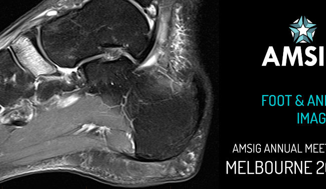
AMSIG Foot And Ankle Imaging Conference
Posted May 16, 2017
During May, members of the Radius team attended the 21st Australasian Musculoskeletal Imaging Group (AMSIG) Annual Scientific Meeting in Melbourne. The group provides a forum for the professional development of musculoskeletal imaging in Australia and New Zealand, as well as promoting education in musculoskeletal imaging, liaison with other similar groups internationally and representation on various government bodies. The conference brought together over 300 attendees, […]
Read More
Education Weekend @ Radius – Ultrasound with Stephen Bird
Posted May 16, 2017
The first weekend in May the Radius sonography group held an ultrasound workshop with Stephen Bird. Stephen is a general sonographer with 25 years of clinical experience, and a very passionate educator of sonography who regularly presents at national and international conferences. Stephen holds a General and a Vascular Diploma of Medical Ultrasound (DMU) and a Masters of Medical Sonography from the University of South […]
Read More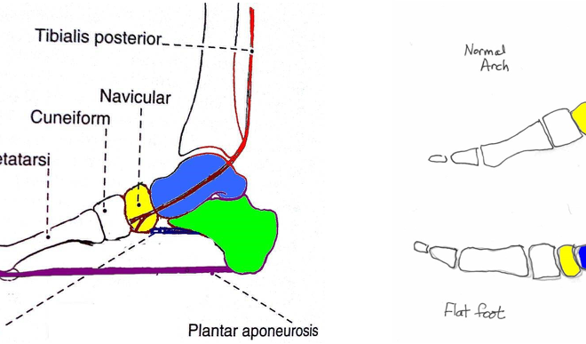
MRI NEWSLETTER: The Spring Ligament, PTT Tear and Adult Acquired Flatfoot Deformity on MRI
Posted May 12, 2017
The posterior tibial tendon is the primary stabilizer of the foot. Other stabilizers include the spring ligament, the tarsal sinus ligaments and plantar fascia. The spring ligament complex consists of the tibio-spring component of the deltoid ligament and the calcaneonavicular ligament , which limits plantar flexion of the talus and stabilizes the talocalcaneal navicular joint. The tarsal sinus ligaments (cervical and interosseous ligaments) limit […]
Read More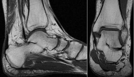
MRI NEWSLETTER: Tarsal Coalition on MR
Posted April 20, 2017
Tarsal coalition is a congenital anomaly with fusion of the tarsal bones. The fusion may be bony, fibrous or cartilaginous. The most common tarsal coalitions are the calcaneonavicular and the talocalcaneal coalition. Tarsal coalitions often lead to painful foot, frequent ankle sprain, flat foot deformity and hindfoot valgus. This may lead to peroneal spastic flat foot. Calcaneonavicular coalition ossifies early and develops symptoms at […]
Read More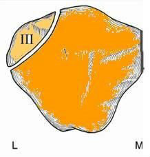
MRI NEWSLETTER: MR of the Bipartite Patella
Posted April 10, 2017
The bipartite patella is a normal patellar variant secondary to failure of fusion. Bipartite patella is often confused with a patellar fracture. Bipartite patella occurs in 8% of the population, is much more common in males 9:1, and bilateral in 50-80%. Bipartite patella occur most commonly in the superior- lateral location, 75% of cases. Bipartite patella is usually asymptomatic and found incidentally with only 2% […]
Read More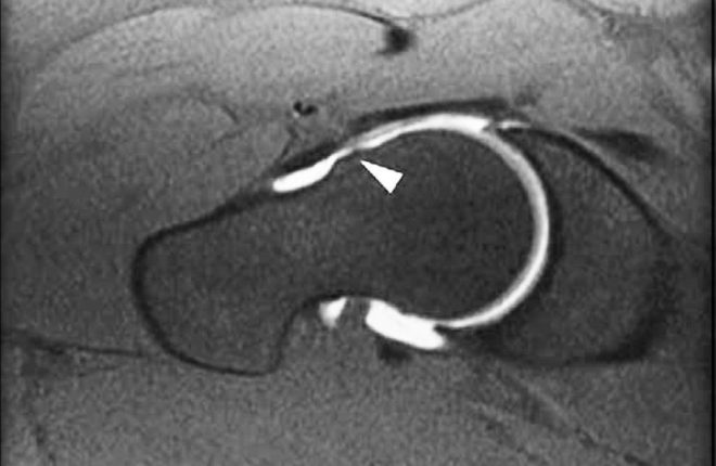
MRI NEWSLETTER: High Incidence of CAM FAI in Athletes
Posted April 4, 2017
Recent studies of FAI reveal the very high incidence of cam deformity (femoral neck bump) in Athletes. The cam deformity is a major risk factor for osteoarthritis of the hip and labral tears. More than half of the athletes studied, average age 25, had evidence of cam deformity as compared to only 23% of age-matched controls. In one study, cam deformities gradually developed during […]
Read More